Methods and techniques
We accept your wishes, approach the treatment with care and always want to choose only methods absolutely essential to the further outcome you seek.
The dividing spindle vizualization method
The combination of the ICSI method with the dividing spindle vizualization method brings other unique possibilities to increase the therapy success further.
Visualization of the dividing spindle of the oocyte is a non-invasive method, thanks to which it is possible to determine whether the egg is mature and fully ready for fertilization.
The maturity of an oocyte is necessary for its successful fertilization. The embryologist usually evaluates as a mature egg that has secreted the so-called pole body and verifies this fact in a “ light” polarization microscope.
However, the oocyte is mature only when the dividing spindle is present inside. It is a tiny structure inside the oocyte that is necessary for the proper distribution of genetic information during cell division. To see the dividing spindle in the cell, it is necessary to look at the oocyte by special polarizing microscope.
In this microscope, the oocyte may look mature but in reality, we can use a polarizing microscope to find out that the dividing spindle is absent, and the oocyte is not yet fully ready for fertilization.
By displaying the dividing spindle, we can then time the fertilization using the ICSI method until the oocyte is ready for fertilization and the probability of its fertilization is the highest. At the same time, if we know the position of the dividing spindle in the oocyte, we can avoid the place where the spindle is located during ICSI and thus avoid irreversible damage to the egg during sperm injection.
Visualization of the dividing spindle using polarization microscopy thus allows us to introduce sperm at the right time to the right place and thus increases the number of fertilized oocytes. It is an ideal solution for couples where the number of collected mature oocytes is low or in previous treatment, for unknown reasons, there was low or zero fertilization.
Embryoscope Plus
If you want to have the best cultivation conditions for your embryos, choose the cultivation in the EmbryoScope +.
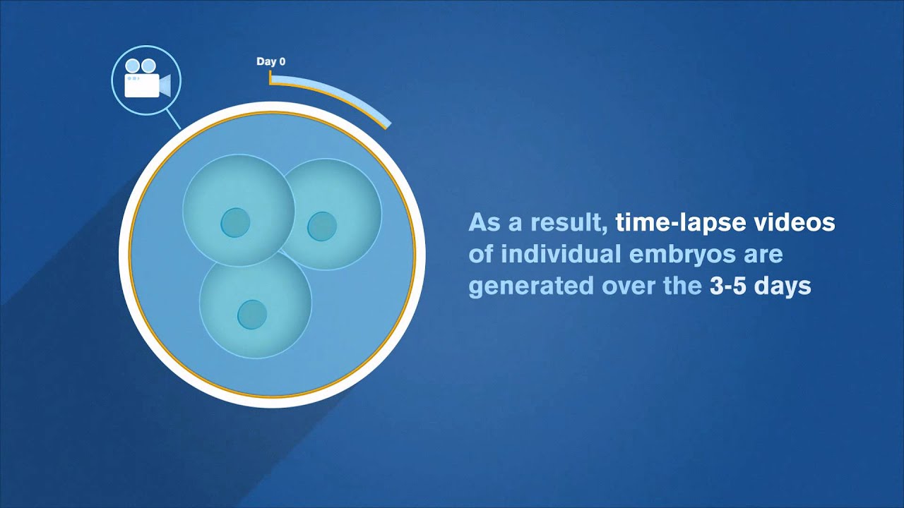
For the successful development of embryos in the laboratory, the stability of the conditions is of the most importance. With the development of new technologies, the old system of cultivation of embryos is replaced by cultivation in so-called time-lapse systems that closely monitor and record the development of embryos without the need to be removed from the ideal cultivation conditions of the incubator.
Our clinic is one of the first in the Czech Republic which have a new EmbryoScope + incubator, which has up to twice its capacity if compared to other time-lapse systems. So we are able to provide the best cultivation conditions for the embryos of all our patients. In addition, EmbryoScope + is designed for separate cultivation – each single-bar coded culture dish contains embryos of one couple and is isolated from another embryos so that when manipulating with one dish, embryo cultivation of other patients in the system is not impaired. In addition, a detailed record of embryo development allows our embryologists enough time and information to select the embryo with the highest development potential.
Embryoglue
EmbryoGlue has the basic composition of a rich blastocyst culture medium and contains a high concentration of hyaluronan and recombinant human albumin. It is uniquely developed to mimic the conditions in the female uterus in order to help embryos implant after transfer.
Simply put – EmbryoGlue helps to glue the fertilized egg to the uterine wall. EmbryoGlue demonstrably increases chances of getting pregnant.
Selection methods of the “best sperm”
a) PICSI (preselected intracytoplasmatic sperm injection)
The so-called pre-selection method involves putting the sperm on a surface similar to the surface of the egg. They are observed and those with the greatest “adherence to this surface” are selected for fertilisation. Their fertilisation potential is considered to be the best.
b) MACS test
Selection of live sperm with undamaged DNA using magnetic nanoparticles. This method ensures that the damaged sperm is not used for fertilising the egg/s.
Sperm with damaged DNA may be capable of fertilising the egg but the embryo development may not be quite optimal. The sperm DNA damage is not visible. It cannot be determined by the shape or properties of the sperm in the microscope even when high magnification is used.
However, the MACS method can discover sperm which contain such damaged DNA. What is more, it can select these sperm and separate them from the rest. We then use only the sperm with undamaged DNA for the fertilisation, or in other words sperm with a significantly higher chance of fertilising the egg, starting the embryotic development and the whole course of the pregnancy.
c) Fertile Microfluidic Sperm Sorting Chip
Chemical-free (does not require any previous processing of sperm sample or use of devices that increase oxidative stress in sperm) method of sorting sperm using a disposable chip.
The method is based on the principle of natural sperm selection in a passage through micro-barriers imitating natural environment of female reproductive system. Selected sperm cells have better morphology, genetic quality and more than double viability and motility than unsorted sperm.
The sperm sample is pipetted into the inlet where the present sperm must actively migrate in the outlet against the movement of the fluid as well as through the above-mentioned micro-barriers. Sperm cells are sorted by separating healthy motile sperm in the outlet from poor-quality sperm captured in the duct.